Nuclear magnetic resonance imaging of the central nervous system
Activities
High-resolution imaging techniques and multinuclear spectroscopy are being developed and validated with a 7T magnet:
- Optimized NMR sequences for rodents and primates (T1, T2 and T2* weighting).
- Optimized NMR sequences for 1H, 13C and 31P cerebral spectroscopy in rodents and primates, automated quantification of short-echo time spectra.
- ZTE (Zero Echo Time) sequence for oxygen-17 imaging.
- CEST ("Chemical Exchange Saturation Transfer") imaging of glutamate in rodents.
- Diffusion-weighted NMR spectroscopy sequences
- Other sequences can be optimized in the framework of collaborative projects.
equipment
Examinations can be carried out on rodents and primates in biosafety level 1, 2 or 3 confinement. The magnet is sealed in a hermetic sarcophagus, making it possible to carry out maintenance interventions on the magnet in confinement conditions.
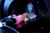
A magnetic resonance imaging experiment on the 11.7Tesla preclinical MRI machine at MIRCen (courtesy of P. Stroppa/CEA).
- Horizontal 7 T magnet (Bruker) dedicated to primates, with radiofrequency coils optimized for the brain and spinal cord of primates.
-
Horizontal 11.7 T magnet (Bruker) for imaging in rodents (funded by
NeuraTRIS) equipped with a cryoprobe.
PET and Radiochemistry
Activities
The expertise of MIRCen in the domain of PET imaging covers the following strategic axes:
1. The
validation of radiotracers in validated animal models of human diseases. |
2. The
validation of animal models by PET with validated radiotracers. |
| 3. The
evaluation of new therapies with a view to transfer to clinical research. |
The PET platform also works closely with the NMR and histology platforms for the three-dimensional reconstruction of multimodal images (Image Processing platform).
PET imaging system for rodents
equipment
The PET platform currently houses:
- Two PET systems are available allowing two PET examinations to be carried out simultaneously for studies in rodents and primates (FOCUS 220, Siemens, Knoxville, TN). These two systems have the following characteristics:
- An axial field of view of 7.6 cm,
- A transaxial field of view of 19 cm,
- A spatial resolution of 1.4 mm,
- A sensitivity of 3.4%
-
Two Swisstrace systems to automatically measure the input function of whole blood
-
Anesthesia and monitoring equipements for small animals
-
A lab dedicated to autoradiographic studies with long half-time isotopes
|
Radiochemistry
Activities
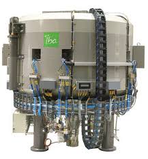
The radiochemistry platform allows the radiosynthesis and the development of new radiotracers specific to biological targets involved in a pathological process. Labeling concerns little molecules (molecular weight < 500 Da) and macromolecules (molecular weight > 500 Da) also. The future in house cyclotron will produce the 2 short half-life radioisotopes
11C and
18F (20.4 and 109.8 minutes respectively):
- For [18F]fluorine labeling, nucleophilic reactions based on [18F]fluoride are used.
- For [11C]carbon labeling, [11C]carbon dioxide is the primary precursor; O- and
N-methylation reactions (involving [11C]CH3I or [11C]CH3OTf) or carbonylation reactions are mainly used.
Another radioisotope with a longer radioactive half-life will be used latter when the pharmacokinetics of the radiotracer and the biological process under study will require it.
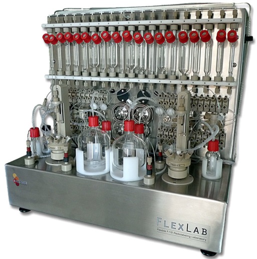
At the end of the radiosynthesis, all radiotracers are analyzed by HPLC in a quality control lab to check their chemistry and radiochemistry purities.
equipment
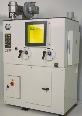
Since the opening of MIRCen, PET tracers have been radiolabeled at the Radiochemistry Laboratory of Frédéric Joliot Hospital, with a biomedical cyclotron and dedicated synthesizers housed in ventilated, shielded areas, according to current regulations. The future new radiochemistry infrastructure will make use of the same facilities.
| 
|
Cyclone 18/18 IBA cyclotron
| Lemer Pax hot cell
|
| |
|
|
Flexlab iphase synthetizer
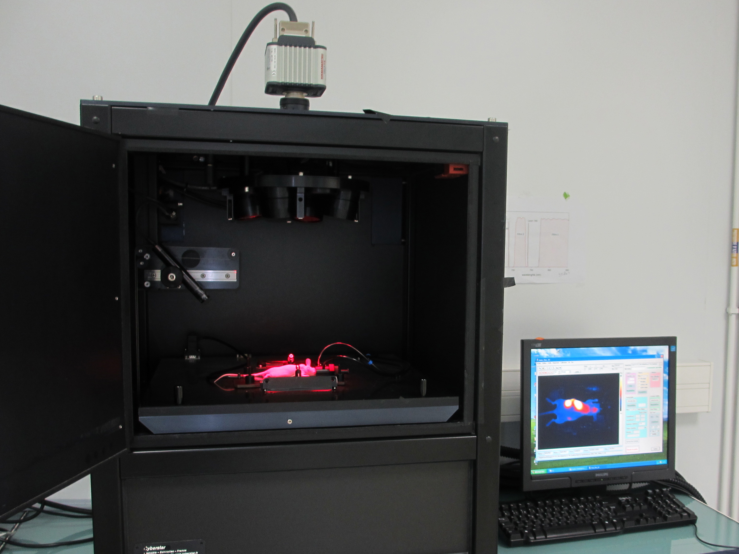
Optical imaging
ACTIVITIES
- Biodistribution of macromolecules (aptamers, antibodies, peptides, nanoparticles)
-
In vivo monitoring of cell migration, differentiation and amplification
-
In vivo imaging of gene expression.
EQUIPMENT
- A near-infrared fluorescence imaging system (fDOT) equipped with two lasers (680 and 740nm) allowing
in vivo 3D quantitative imaging at a resolution of mm3..
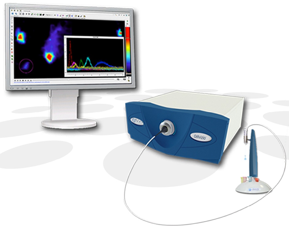
- A confocal fiber endo-microscopy system (pCLE) equipped with a 488nm laser and allowing the in vivo imaging of probes or fluorescent proteins at a resolution of a few microns.
| |
|
 |